
CELL LINE Development
Category
1. Isolasi DNA/RNA
(Step description Isolasi DNA/RNA)
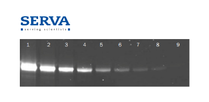
SERVA Triple Color Protein Standard II
Cat. No: 39257.01

SERVA Triple Color Protein Standard II is a mixture of 10 pre-stained proteins of a molecular weight range from 11 to 180 kDa (separation on a SDS Tris-Glycine gel). Proteins are covalently coupled with a blue chromophore except for one green band at 25 kDa and one red band at 75 kDa (separation on a SDS Tris-glycine gel).
SERVA PRiME Lightning Red
Cat. No: 43402.01
SERVA PRiME™ Lightning Red is a fluorescent dye for rapid labeling of proteins prior to SDS PAGE, making any staining and washing steps after electrophoresis unnecessary. In addition, the dye is fully compatible with mass spectrometry and other downstream methods like Western Blotting.
Quick Coomassie® Stain
Cat. No: 35081.01
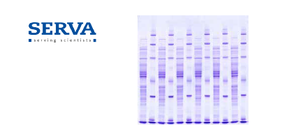
Quick Coomassie® is a fast and non-toxic 1-step Coomassie® stain that is up to 50 times more sensitive than other quick stains. To detect electrophoretically separated proteins, colorimetric staining methods are common. Coomassie® and silver are mainly used. The best method for a specific application strongly depends on the detection limit, the compatibility with downstream applications and detection instruments.
Acrylamide/Bis Solution, 37.5:1 (40 % w/v), 2.6 % C (500 ml)
Cat. No: 10681.01
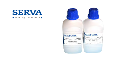
SERVA’s ready-to-use acrylamide/Bis solutions are suitable for all protein electrophoresis techniques – from standard SDS PAGE to high performance horizontal 2D electrophoresis. For manufacturing of various polyacrylamide gels SERVA has developed a profound knowledge base in making acrylamide and buffer solutions
Laemmli Buffer 10x, for SDS PAGE (2L)
Cat. No: 42556.01
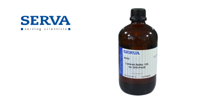
To obtain optimal protein separation in vertical SDS PAGE it is crucial to use the appropriate buffer system. The standard Tris/Glycine (Laemmli) buffer system is suitable for the MW range 7 – 200 kDa. Supplied as 10 x concentrate. Contains 0.25 M Tris, 1.92 M glycine and 1 % SDS in aqueous solution.
2D HPE™ Large Gel NF 12.5 % Kit
Cat. No: 43304.01
2D HPE™ large gels are suitable for running 1 x 24 cm IPG strip (e.g. SERVA IPG BlueStrip) plus one marker lane by horizontal electrophoresis on HPE™ Tower, HPE™ BlueHorizon™ or Multiphor II™. 2D HPE™ large gels are available on standard backing or on non-fluorescence (NF) backing for fluorescence staining and labelling. All kits include 4 gels, running and equilibration buffers, wicks and cooling contact fluid.
2D Gel DALTsix NF 12.5 % Kit
Cat. No: 43313.0
The 2D Gels DALT Kits contain 6 or 12 gels for Ettan DALTsix and Ettan DALTtwelve, respectively plus buffer kit. Buffer kits contain anodal and cathodal buffers as well as IPG equilibration buffer and agarose for strip fixation.
SERVAGel™ TG PRiME 12 %, 2D well (10 gels)
Cat. No: 43268.01
Obtained from proprietary development, the precast gel SERVAGel™ TG PRiME™ 12 % features an extended shelf life and short electrophoresis times by using a standard Tris/glycine buffer system. The 2D gel has one very planar slot for optimum transfer of proteins in the second dimension. For the first dimension IPG strips of 7 cm length can be used. The separation distance is 7 cm.
SERVA IPG BlueStrip 3-10 (7 cm)
Cat. No: 43001.01

SERVA IPG BlueStrips are dried gel strips with immobilized pH-gradient used in high resolution 2D-gelelectrophoresis of proteins. The homogeneous polyacrylamide gel matrix is covalently bound to GEL-FIX™ to stabilize the gel. Additionally, a non-binding cover film (GEL-FIX™ for Covers) protects the gel from damage and contamination.
SERVA Proteome Markers
Cat. No: 39220.01
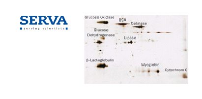
SERVA offers the unique set of Proteome Markers containing 8 proteins qualified for 2D gel electrophoresis and application in liquid chromatography/ mass spectrometry. Proteins ranging from 11.7 to 77 kDa and spanning the entire pI range are supplied in equimolar amounts.
SERVA HPE™ Coomassie Staining Kit
Cat. No: 43396.01
Colloidal staining kit for highly sensitive staining of 1D and 2D gels after electrophoresis. The kit contains two components to be mixed together prior to use. Kit contains enough reagents to stain 4 large 2D HPE™ gels. Reagents are MS compatible for downstream mass spectrometry analysis.
2. Gene Splicing
(Step description Gene Splicing)
HPE™ BlueHorizon™ System
Cat. No: HPE-BHSYS.01
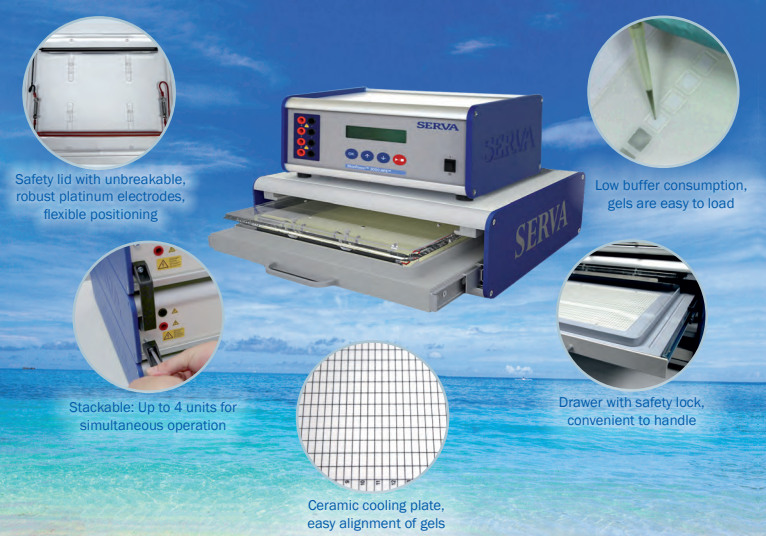
The HPE™ BlueHorizon™ is a flatbed system for horizontal electrophoresis using precast gels, self-cast gels and gel strips. Main applications are isoelectric focusing (IEF - like CSF, seed and food analysis, EPO, separation of antibodies and other recombinant proteins), SDS and native PAGE, 2D electrophoresis as well as separation of nucleic acids. The HPE™ BlueHorizon™ flatbed electrophoresis system provides unmatched resolution, reproducibility and sensitivity – the first true "High Performance Electrophoresis (HPE)“ system.
HPE™ BlueTower System
Cat. No: HPE-TS2.01
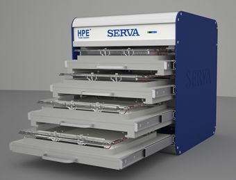
The HPE™ BlueTower System allows electrophoretical separations in up to four horizontal gels at the same time. It is used for 1D and 2D electrophoresis gels, where multiple runs are an important demand. Structurally, the HPE™ BlueTower consists of four horizontal electrophoresis chambers, which are built as movable drawers into a metal housing.
BlueVertical™ PRiME™
Cat. No: BV-104.01
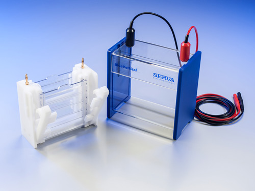
The BlueVertical™ PRiME™ is a dual mini tank system to operate one or two precast gels. It accomodates SERVAGel™ TG PRiME™, all other types of SERVAGel™ and all other commercially available precast gels with an outer cassette dimension of 10 x 10 x 0.7 cm. Separation of proteins by SDS PAGE, native PAGE and IEF can be carried out as well as separation of nucleic acids.
3. Vector Insertion
(Step description Vector Insertion)
Immobilon™-P-membrane
Cat. No: 42581.01
Immobilon™-P-membranes developed by Millipore Corp. are specially designed for Western Blot techniques. The membranes, made of polyvinylidenfluoride (PVDF), show excellent mechanical stability and are compatible with most staining procedures including immunological methods.
Blotting Paper for Western Blotting
Cat. No: AC2111
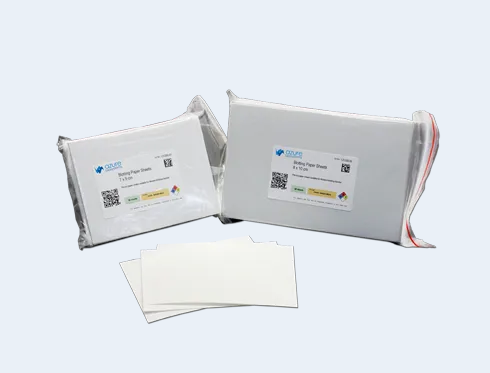
Pre-cut blotting papers save time and reduce waste for the easiest Western blotting workflow. Forget about measuring and cutting blotting paper to the size of your mini-gels. Use these pre-cut papers to assemble your transfer “sandwiches”.
Plasma Membrane Fraction Western Blot Cocktail
Cat. No: ab139413
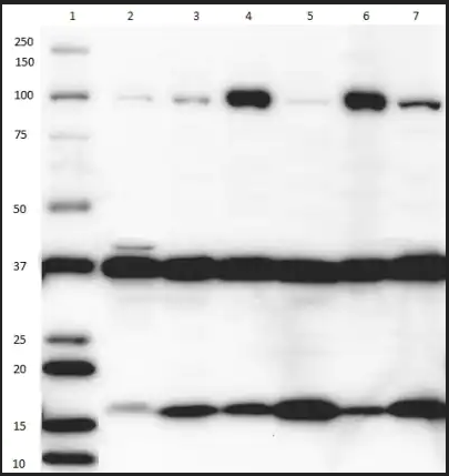
ab139413 contains 3 mouse monoclonal antibodies each targeting a specific organelle marker. The presence of plasma membrane is determined by Anti-Sodium Potassium ATPase antibody; cytosol by Anti-GAPDH; and nucleus by Anti-Histone H3 (di methyl K9). This cocktail is suitable for determining the purity of organelle isolates prior to further characterization. This product is particularly valuable to researchers working in organelle proteomics.
BlueBlot Semi-Dry Blotter SD
Cat. No: BB-SD11
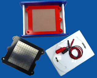
The BlueBlot™ Semi-Dry Blotter forms a homogeneous electrical field that guarantees fast and efficient transfer of proteins from gel to membrane.
4. Transfer into Host Cell
(Step description Transfer into Host Cell)
Azure 600, The Ultimate Western Blot Imaging System
Cat. No: Azure 600
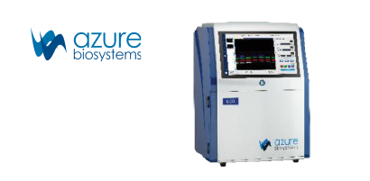
The two-channel NIR Fluorescence detection enables sensitive multiplex Western Blotting with minimal background. Image and quantify two different targets in the same position more efficiently using NIR, and normalize to fluorescent total protein stain or a housekeeping protein in the green channel without needing to strip and re-probe your WBs.
Azure 200, Gel Imaging Workstation
Cat. No: Azure 200

Easily image DNA and protein gels images without a darkroom with the Azure 200. Use your choice of dyes or stains and the system automatically selects the light source and filters for you—UV for Ethidium bromide-stained DNA gels, blue light for SYBR®Safe or similar dyes, white light for Silver stain or Coumassie Blue.
AzureSpot Analysis Software, Advanced Analysis for 1D Gels and Blots
Cat. No: AzureSpot Analysis Software
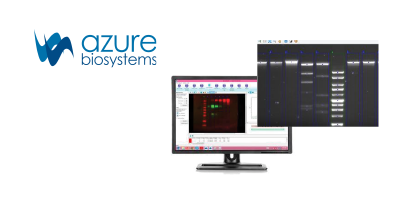
AzureSpot helps you interpret your data easily and accurately. AzureSpot can automatically detect lanes and bands, even on distorted gels, and apply your selected method of background correction. Visualize intensity plots by lane (sample) and channel (probe) to get detailed information about your proteins of interest.
5. Growing Host Cell Media
(Step description Growing Host Cell Media)
Workflow Protein Tech Lainnya
Upstream Workflow
Produksi biosimilar atau active pharmaceutical ingredients (API) dari biofarmasetikal dimulai dari pembuatan API menggunakan sel hidup yang dikenal dengan proses upstream.
Downstream Workflow
Setelah peneliti selesai dengan proses upstream. Kini yang dilakukan adalah proses downstream, yaitu proses pemurnian dari culture media menjadi Proses downstream dilakukan setelah proses upstream.
Microbial Identification
There is a Biolog System for any size laboratory, from full and semi-automatic to manually read systems. with 20-years more experience serving customer.
Sampaikan Kebutuhan Riset Anda
Punya pertanyaan atau permintaan khusus seputar produk dan layanan Protein Tech? Hubungi tim kami: